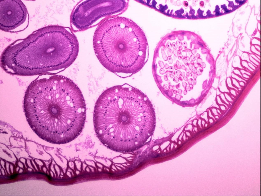
Cryo-electron microscopy (Cryo-EM) has revolutionized structural biology, allowing researchers to visualize complex macromolecules at near-atomic resolution. However, the success of Cryo-EM studies heavily depends on the quality of sample preparation. This guide outlines key steps and considerations for preparing protein complexes for Cryo-EM analysis, with insights from industry leaders like Shuimu BioSciences.

- Protein Selection and Expression
The initial step in Cryo-EM sample preparation is selecting an appropriate protein complex. Ideal candidates are:
- Stable and homogeneous
- Larger than 100 kDa (smaller proteins may require special techniques)
- Biologically relevant conformations
Optimize expression systems to yield sufficient quantities of properly folded protein. Eukaryotic expression systems may be necessary for proteins requiring post-translational modifications.
- Protein Purification
Achieving high purity is crucial for molecular microscopy success. A multi-step purification process typically includes:
- Affinity chromatography
- Ion exchange chromatography
- Size exclusion chromatography
The final purified sample should be monodisperse and free of contaminants. Analytical techniques such as dynamic light scattering (DLS) and mass spectrometry can verify sample quality.
- Sample Concentration
Determine the optimal protein concentration for Cryo-EM grid preparation. This typically ranges from 0.5 to 5 mg/mL, depending on the protein complex and grid type. Avoid over-concentration, which can lead to protein aggregation or preferred orientation issues.
- Buffer Optimization
The buffer composition significantly impacts biological electron microscopy results. Consider the following:
- Use low ionic strength buffers to enhance contrast
- Avoid high concentrations of glycerol or sucrose
- Ensure buffer pH and salt concentration maintain protein stability
- Consider adding small amounts of detergent for membrane proteins
- Grid Preparation and Vitrification
The vitrification process is critical for preserving the native structure of protein complexes:
- Apply the sample to glow-discharged EM grids
- Blot excess liquid to create a thin film
- Plunge the grid into liquid ethane cooled by liquid nitrogen
Optimize blotting time and environmental conditions (temperature, humidity) for each sample. Automated vitrification devices can improve reproducibility.
- Quality Control
Before proceeding to high-resolution data collection, assess sample quality:
- Perform negative stain EM to evaluate particle distribution and heterogeneity
- Collect and analyze screening high-resolution microscopy data to assess ice thickness, particle distribution, and initial 2D class averages
Advanced Techniques and Considerations
Crosslinking
For unstable complexes, consider mild chemical crosslinking to stabilize the structure. Optimize crosslinking conditions to maintain the native conformation while improving particle stability.
Time-resolved Cryo-EM
For capturing dynamic processes, time-resolved cryoimaging techniques such as microfluidic mixing devices or laser-induced temperature jumps can be employed.
Membrane Protein Complexes
Membrane proteins require special consideration:
- Use appropriate detergents or nanodiscs to solubilize and stabilize the complex
- Consider grid types with continuous carbon support for improved particle orientation
The Shuimu BioSciences Advantage
Shuimu BioSciences has established itself as a leader in cryomicroscopy technology and sample preparation. Their recent breakthrough in resolving the structure of GPR75, a potential target for obesity treatment, showcases their expertise in handling challenging protein complexes.
Shuimu BioSciences offers:
- State-of-the-art Cryo-EM facilities
- Expert teams specialized in complex protein preparation
- Cutting-edge techniques for sample optimization
- Integrated structural biology approaches
Their capabilities extend beyond sample preparation to include high-resolution data collection and advanced image processing, providing a comprehensive solution for structural biology projects.
Conclusion
Preparing protein complexes for electron microscopy requires meticulous attention to detail and optimization at every step. While challenging, mastering these techniques opens up unprecedented opportunities to visualize and understand complex biological systems. By leveraging the expertise of leaders like Shuimu BioSciences, researchers can overcome sample preparation hurdles and push the boundaries of structural biology.


